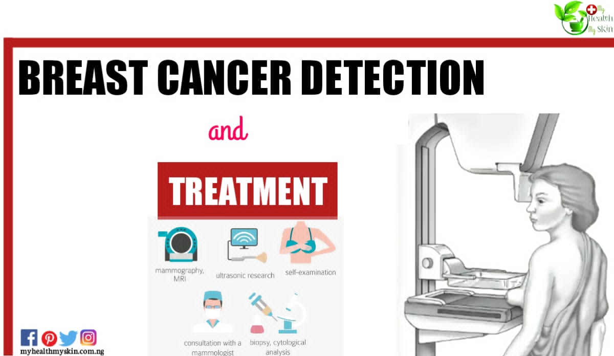
Breast cancer is a condition in which breast cells grow uncontrollably. There are several types of breast cancer (carcinoma). The type of carcinoma depends on the breast cells that develop into cancer.
Breast cancer can start in different parts of the breast. The breast consists of three main parts: ducts, lobes and gastric tissus.
The lobes (pores) are the milk producing glands, the ducts on the other hand, are the tubes that carry the milk to the nipple while the gastric tissues (made up of fibrous and fatty tissue) surrounds and holds everything together. Most breast cancers start inside the pores or lobules.
Breast cancer can spread beyond the breast via the blood and lymphatic vessels. When the carcinoma spreads to other parts of the body, it is called metastasis.
Types of Carcinoma
The most common types of carcinoma are –
Infiltrating ductal carcinoma: Cancer cells grow outside the ducts and in other parts of the breast. The invasive cancer cells may spread, or metastasise, to other parts of the body.
Infiltrating lobular carcinoma:
The cancer cells spread from the lobules into the accessible breast tissue. They can also spread to other parts of the body, which makes them invasive.
There are other, less common types of carcinoma such as Paget’s disease, medullary carcinoma, mucinous carcinoma and inflammatory carcinoma.
One known causal disease of breast carcinoma is Ductal carcinoma in situ (DCIS). In this case, the cancer cells are only inside the ducts and have not spread to other breast tissue.
Methods of Breast Cancer Detection: breast cancer♋ screening
Breast cancer screening helps to detect cancer at an early or asymptomatic stage. In many cases, screening is done when symptoms have already appeared, making treatment and prognosis more difficult.
Cancer screening can be started if you have several risk factors or, depending on your age, ideally after 35-40 years.
Mammography
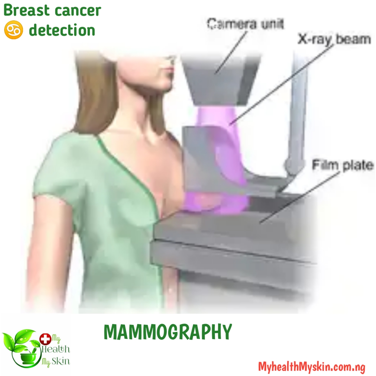
A mammogram is an imaging test that allows radiologists (doctors who specialise in radiology) to see abnormalities or changes in the breast tissue. It is the only breast cancer ♋ detection method that has been shown to reduce breast cancer mortality by 40%.
Why do I need a Mammogram?
Mammography usually detects breast cancer at an early stage, when it is small or even when it is not visible or hardly visible. At this time it is easier to treat.
Types of Mammography
Screening mammography: used to look for signs of breast cancer in women who have no obvious masses or symptoms. Two X-ray slides from different angles are needed for each breast.
Diagnostic Mammography: this breast cancer detection method can be used to examine the breast specifically to determine the presence of tumour growth.
They can also be used to assess women undergoing treatment for breast cancer.
How do mammograms work?
A mammogram is a machine designed solely for the examination of breast tissue. This machine receives low-dose X-rays that penetrate the tissue, making it easier to detect any abnormalities.
This machine has two plates, an upper plate and a lower plate, which help compress the breast, causing the tissue to separate, giving a better image.
There are now new mammography machines called breast tomosynthesis or 3D mammography. The breast is squeezed once, the machine moves across the breast and the machine takes several low-dose X-rays, which are then combined by a computer into a 3D image.
This type of mammogram allows the doctor to see more clearly in very dense tissue lesions that may be hidden in the overlapping tissue of traditional mammography, thus avoiding additional diagnostic or follow-up sessions.
How to prepare for a mammogram?
Go to a diagnostic imaging laboratory where mammograms are performed and the staff are experienced in performing and interpreting mammograms.
Try to visit the same facility each time so that your mammograms are on file.
If you go to a new location, keep a list of previous mammogram locations and dates.
Make an appointment if you have problems with your breasts, such as soreness or swelling.
Do not use deodorants or antiperspirants during the mammogram – they contain substances that can look like white spots. You can use deodorant after the mammogram.
You will be asked to leave your breasts uncovered; comfortable clothing is recommended.
Ask about any recent problems or changes you have noticed before your mammogram.
Ultrasound or Breast Ult
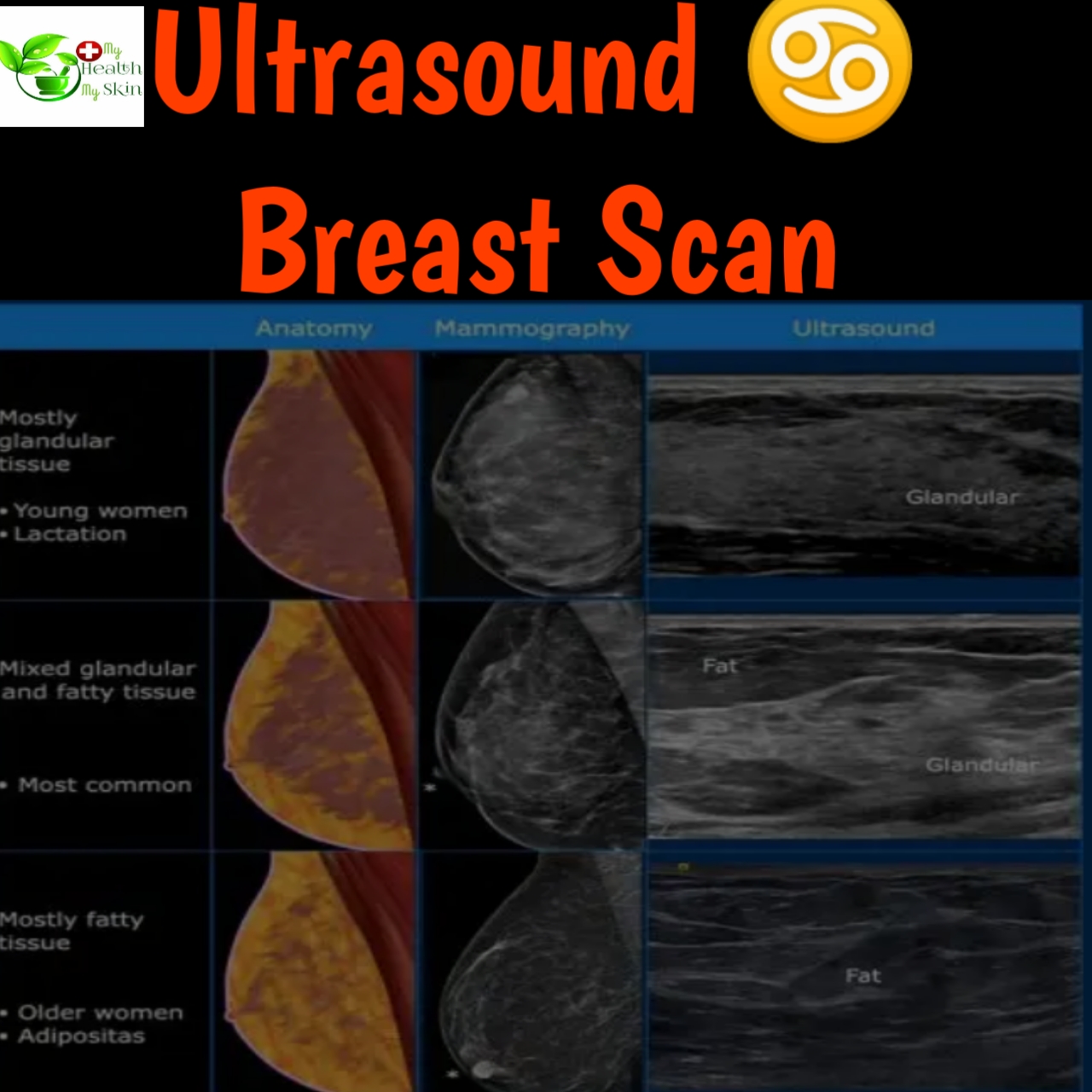
A breast ultrasound is used to assess breast tissue and to see if a lump is a cyst or a solid tumour. It can also be used to see changes that have occurred during a mammogram.
It is the method of choice for women under 40 years of age.
Ultrasound can be very useful to distinguish between fluid-filled cysts and solid masses (the latter require further investigation to be sure they are malignant).
Breast ultrasound can also be used to take a biopsy to look for cancerous cells in the breast. It can also be useful for lymph nodes under the arm.
Ultrasound as a breast cancer detection method is an instrument that is easier to find and does not expose you to radiation and is cheaper.
How is it done?
An ultrasound creates an interior image of the mamary gland (breast) through the use of sound waves.
A gel is applied to the breast, a transducer emits sound waves and receives the echo reflected from the tissue, creating an image on a computer. It is usually painless, but pressure must be applied to the breast.
Magnetic Resonance Imaging
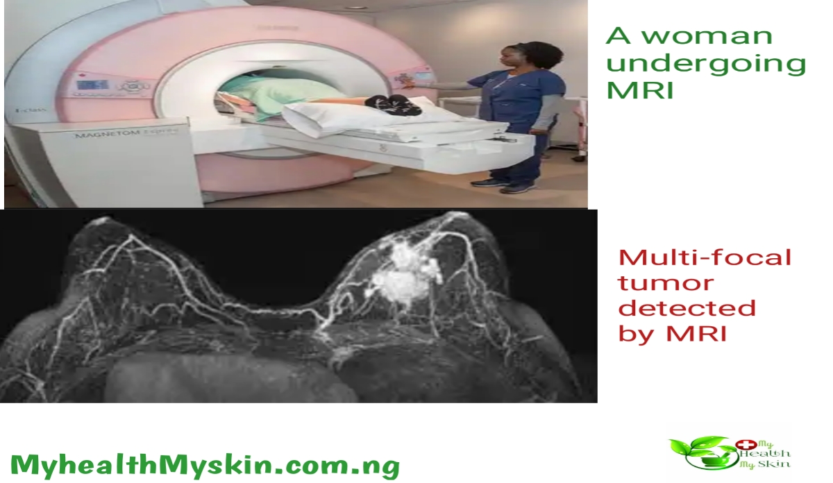
Another type of breast cancer detection method is the MRI. The breast MRI uses radio waves and powerful magnets to produce detailed images of the inside of the breast.
When is the breast MRI used?
MRI is usually used in women who have already been diagnosed with breast cancer and helps measure the size of the cancer and assess its growth rate, as well as detecting possible tumours in the opposite breast, or when mammography and ultrasound are not enough to diagnose cancer.
Although MRI can detect many types of cancer that are not easily seen with mammography, it is possible to detect or find something that is not cancer, so-called “false positives” that should be evaluated to rule out the possibility of cancer.
Such situations require further investigation or biopsy.
It is also the most sensitive method of assessing the integrity of implants.
What do I need to know about an MRI scan?
An MRI scan requires special equipment. Not all diagnostic centres have special equipment for breast MRI.
The magnetic resonance imaging (MRI) takes pictures from different angles, allowing you to see soft parts of the body that are sometimes difficult to see with other imaging methods.
Tips for getting an MRI
Visit a reliable health centre with staff who are experienced in performing and interpreting breast MRI scans.
If you have a phobia of enclosed spaces for any reason, you may need to take medication to help you relax during the scan.
Note: REMOVE ANY METAL OBJECTS.
It is advisable to remove all metal objects and ask to undress and put on a gown for the scan.
You should tell the technician if there are any metal objects on your body. In general, there are metals that do not affect you and others that do, examples of such harmful metals are defibrillators (also called pacemakers), brain aneurysm clips, implants, metal vascular coils, etc.
Breast Biopsy

If other tests show suspicious lesions, a biopsy may be needed. It does not necessarily mean that you have cancer, it is for further clarifications.
Most results are not cancerous, but a biopsy is the only way to find out. During a biopsy, the doctor takes tissue from the suspected area for examination in a laboratory to confirm or rule out the presence of cancer cells.
Types of breast biopsy
Some types of biopsy use a needle, others use an incision. Each type has its own advantages and disadvantages. The type depends on several factors:
How suspicious the breast change looks.
How suspicious is it?
In what part of the breast is it located?
What kind of damage.
How severe the lesion is.
Personal preference.
We recommend that you ask your doctor what type of biopsy he or she will do and what you can expect before and after the examination.
Biopsy Methods:
Fine needle aspiration (FNA) biopsy.
Nuclear bladder biopsy
Surgical (open) biopsy
Lymph node biopsy (open biopsy).
Breast Exam
This examination includes palpation of the breasts and mammary glands for unusual lumps or bumps.
This examination can be carried out by a specialist or by yourself.
This type of examination is also called a self-examination. Periodic self-examination can help detect changes in your breasts.
It is important to remember that these tests do not prevent breast cancer or reduce your risk, it is important to notice changes and contact your specialist who will tell you what is going on, as it is often confused with other conditions.
You should know that a mammogram is more effective than a self-examination, as is an MRI and breast ultrasound.
Experts disagree on when to perform or start breast cancer screening, it is best to consult your doctor and discuss the risks with him or her, as all bodies are different.
Summary
You now know the most common tests that can help you in breast cancer♋ detection or confirm the onset of cancer.
There are other tests that can help you detect cancer, but they are more specific. Always consult your doctor first.
If you have reached this point, you may find this information interesting or useful. If so, please share it with your acquaintances or the best people you think would benefit from reading this blog. The fight against breast cancer is in your hands.
To stay on top of your game, always have a handful of Information about your health.
Understanding health information determines how well a person receives and utilizes the health information and services available to them, especially in making good health decisions. We can help you by sharing the information you need to make the best decisions about your health. To stay informed, sign up to our weekly publications, it’s free.

